Focus: Separation and Concentration of Microorganisms from Food Matrices
Updated: 1st Nov 2019
Key Points
- Improvements in homogenization instruments and filtration have shortened the enrichment time needed for the detection of pathogens.
- International standard protocols now include Immunomagnetic separation for high-risk pathogens.
- Viable PCR technology can give real-time pathogen detection results for HACCP monitoring.
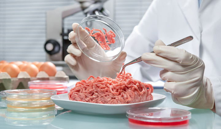
One of the most important tasks undertaken by food microbiology laboratories is the testing of samples for the presence of foodborne pathogens and potential spoilage organisms. Microbiological standards and criteria applying to food often state that pathogens such as Salmonella, E. coli O157, and Listeria monocytogenes should be absent in a specified weight of the sample, usually 25g. Therefore methods must be capable of detecting a single viable cell of the target species in a 25g of the food sample. Traditional culture-based methods have that capability, but take several days to produce a result because of the need to amplify low numbers of cells to levels that can be detected in culture.
Reducing the time needed to complete pathogen detection tests has been a primary goal for food microbiologists for several decades, with much research in developing rapid methods as a result. Now laboratories have an array of detection methods based on technologies like PCR and enzyme immunoassay, which can cut test times considerably and be mostly automated.
The challenge is magnified still further by the complexity of food matrices. Most foods are composed of many different materials, including large proteins, fats and oils, sugars, and complex polysaccharides, often arranged in a complex three-dimensional structure. Many also contain a range of other non-pathogenic microorganisms, sometimes present in high numbers, but poses no health threat to the consumer. The target pathogen cells might be a tiny component of the overall microflora and attached within the food matrix as single cells or clumps of cells. Before rapid detection methods can be used successfully, it is usually necessary to separate the target cells from the food matrix and the background microflora. Once the cells have been separated, further time savings are available if they can then be concentrated to allow faster detection.
Methods
To note: The sample must be suspended in a relatively large volume of liquid - diluent or growth medium - before separation and concentration can be attempted. Virtually all current pathogen detection methods include either an enrichment culture or suspension step.
Enrichment culture
Recent developments: Depending on the pathogen of interest, the ISO and BAM (US, FDA) standards may vary with media used in protocols, but rarely do their enrichment times differ. Some rapid methods reduce the enrichment time in half, and have yet to be included in ISO and BAM standards. The supplier of the rapid method and its potential user will have to obtain validation for these methods with the proper certification bodies.
For example, the pathogen E. coli 0157 has a standard enrichment time of 18 hours in both the ISO and BAM protocols. But with recent advancements in filtration and concentration, food microbiologists can now complete the entire detection protocol in just 8 hours.
International standards: At present, the traditional methods still apply to international standards in detecting low numbers of pathogens in food samples. Many standard methods include a two-stage enrichment culture. The first step, or pre-enrichment, involves suspending the required sample weight (10-25g) in a large (100-250ml) volume of a non-selective medium designed to resuscitate damaged cells and promote microbial growth. After incubation, typically at 37oC for 18-24 hours, a small volume of the culture is transferred to a suitable selective enrichment medium designed to suppress the growth of competing organisms while allowing the target pathogen to multiply. In theory, such methods can amplify a single viable cell to detectable levels, even if it remains attached to the food matrix. There is no need to treat the sample to ensure that microbial cells are freely suspended in the medium. Though simple and reliable, traditional enrichment culture-based methods are extremely time-consuming, often taking several days to complete.
Food sample suspensions
Pathogen detection methods that do not utilize some form of enrichment culture still require the preparation of a sample suspension before separation, concentration, and detection techniques can be applied. Ideally, the initial suspension will be prepared in such a way that each target cell present is freely suspended in the diluent. Most foods will require a homogenization step using a stomacher or a blender. Blenders should not be used in measuring Aw as they can generate heat, causing water present in the food to evaporate and effect Aw readings.
Traditional quantitative methods (e.g., for total viable counts) often specify some form of mechanical agitation to release microbial cells from the sample matrix. Some protocols still include rotary bladed blenders as the preferred method of producing a suspension, but the need to clean and sterilize the blender between samples and problems with heat generation in the suspension limit their effectiveness. Paddle type blenders like the Seward Stomacher® overcome some of these problems by processing the sample in a sterile plastic bag so that the paddles do not make contact with the sample and remain clean. They are also able to handle large volumes of liquid. Paddle blenders are now widely used in food microbiology laboratories and have replaced bladed blenders.
A more recent development is the Pulsifier® from Microgen Bioproducts, which replaces the paddles with a high-speed oscillating ring, producing shock waves as well as agitation. The Pulsifier can release more microbial cells into suspension with less disruption to the food matrix than other blenders, and without creating large quantities of food debris in the suspension. Other methods for preparing initial suspensions, such as sonication, have been investigated, but are not widely used.
Separation and concentration
To note: Once microbial cells have been released from the food matrix into suspension, the target pathogen can be separated, and the cells concentrated for more rapid detection. Many different physical, chemical, and immunological methods have been devised to accomplish this, including ion exchange columns and biphasic partitioning. But three techniques form the basis of most practical applications: membrane filtration, centrifugation, and immunomagnetic separation (IMS).
1. Membrane filtration
Filtration has obvious attractions as a means of separating and concentrating microbial cells from sample suspensions. It is relatively inexpensive and straightforward, with the potential to process large volumes of suspension quickly. Some filtration products on the market can work while the food is being homogenized. This is usually called a pre-filtration step and allows a more advanced filtration membrane to work more efficiently. The InnovaPrep Concentrating Pipette, for instance, will automatically run the diluent through its filtration membrane of which microbes attach to. An elution is produced using proprietary wet foam technology and can now be lysed and run on a PCR thermocycler to fulfill pathogen DNA detection requirements. This whole process, which also includes the enrichment stage, takes only 8 hours.
There are difficulties involved when working with high-fat processed foods when filtering. Food debris can block the membrane and prevent some of the target cells being collected. Some improvements in the filterability of food suspensions can be achieved by techniques such as trypsin digestion or the addition of surfactants, but this may also interfere with pathogen detection and reduce cell viability.
For filterable samples, such as beverages and some dairy products, practical separation and concentration methods based on membrane filtration have been developed and combined with a variety of microbial detection technologies as commercially available instruments and test systems. Examples include the ScanRDI from BioMeriux, which uses a solid-phase cytometry technique to detect viable cells directly on filters. Microscopy-based methods, such as the direct epifluorescent filter technique (DEFT), are also still used in some laboratories to count bacterial cells on membrane filters.
2. Centrifugation
Centrifugation has also been used successfully to separate and concentrate microbial cells in food suspensions, although applications are limited by the practical considerations of processing large volumes of liquid. Low-speed centrifugation can be used to remove food debris while leaving bacterial cells in suspension, and higher speeds can then be used to concentrate bacteria. Alternatively, methods based on density gradient centrifugation can be used. For example, buoyant density centrifugation (BDC) has been used in rapid detection of foodborne pathogens. It separates bacterial cells and food particles with different buoyant densities by flotation and sedimentation and to remove PCR inhibitors. Some commercial applications have also been developed, notably the automated Foss Bactoscan™ FC instrument designed for the dairy industry. This can separate bacterial cells from milk with the aid of a density gradient, before detection and enumeration by flow cytometry.
3. Immunomagnetic separation (IMS)
IMS technology was first developed in the 1980s and is now widely used in food microbiology to separate target pathogens from other microorganisms. Magnetized polystyrene beads or other particles are coated with antibodies specific to the target organism, and these are then added to a volume of the sample directly (where high counts are likely), or more often to a pre-enrichment culture. After a short incubation, the target cells are immobilized on the surface of the beads and can be concentrated into a pellet using a powerful magnetic field. The sample can then be discarded, and the pellet is resuspended and washed to remove food debris and other material. The immobilized pathogens can be detected by culture, or by rapid methods, including PCR and ELISA. IMS is a very effective separation and concentration technique, although it is not suitable for processing large volumes of sample or culture, partly because high concentrations of beads are needed to bind the target cells.
Magnetic beads and particles for food microbiology applications have been available commercially for many years. The best known are Dynabeads®, supplied by ThermoFisher Scientific and designed for selective capture and concentration of a range of foodborne pathogens from pre-enrichment cultures. Dynabeads specific to Salmonella Listeria and several VTEC serotypes are available. Dynabeads can also be used in an automated process using the BeadRetriever™ system. Further development of automated IMS technology is illustrated by the Pathatrix® Auto System, also from Thermo Fisher. Pathatrix is unique in allowing up to ten samples to be pooled after pre-enrichment, providing considerable cost savings through reduced detection testing. The separation and concentration process can be completed within 15 minutes.
Future developments
Although methods and instruments designed to speed up the process of separating and concentrating microorganisms from foods are available, there is still a need for new methods to be developed to support PCR and immunology-based detection tests. At present food microbiologists are still reliant on enrichment cultures to amplify low numbers of pathogens to a detectable level, and the goal of instant detection has yet to be achieved.
One approach to moving away from dependence on enrichment procedures is to combine some of the technologies described above in multi-step methods. For example, a protocol using a combination of filtration, low and high-speed centrifugation and buoyant density gradient centrifugation has been described. It is capable of a 250-fold concentration of target pathogens in food samples and detected 10 CFU/g of Salmonella in chicken in 3 hours when combined with a PCR detection technique. Such a method would not be practical for routine application in a food microbiology laboratory but does indicate the potential for the development of multi-step methods, especially if they can be automated.
Get the latest updates in Rapid Microbiological Test Methods sent to your email? Subscribe to the free rapidmicrobiology eNewsletter


